Fluorescence Microscopy of Brugia
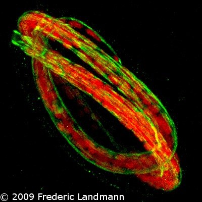
1. B. malayi Pretzel embryo about to hatch, PI staining (red) and actin cortex (Phalloidin in green)
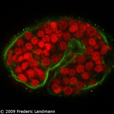
2. B. malayi 1.5 fold embryo, same stainings
3. B. malayi zygote in uterus. In red, the polar body and Wolbachia; in green the actin cortex and anti-acetylated H4 staining the chromatin. Around the zygote in red, sperm cells.
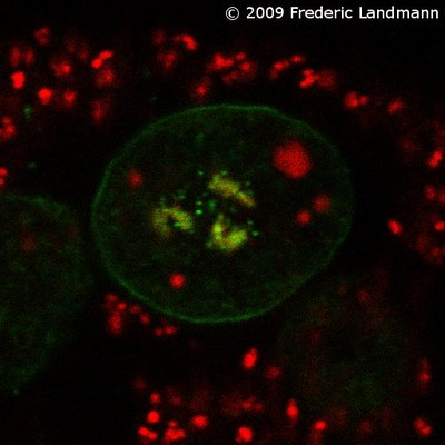
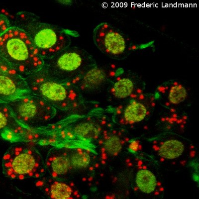
4. Oogonia in ovary, same stainings as above, showing Wolbachia around the nucleus
5. Distal part of the ovary, germline cells filled with Wolbachia (red). In green, the chromatin (acH4) and the central actin-rich rachis.
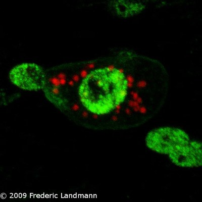
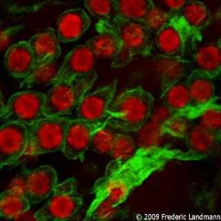
6. Distal part of the testis, same stainings, NO Wolbachia
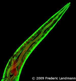
7. male posterior part, actin (green), PI (red) and autofluorescnece of the spicules in red
8. close up of 8
9. female anterior part, actin in green, PI in red, see muscles, pharynx and ovejector.
10. male spicules
11. ovejector, actin
12. ovejector, DNA (PI)
13. Merge of 12 and 13

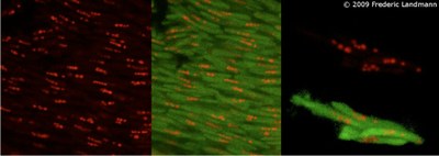
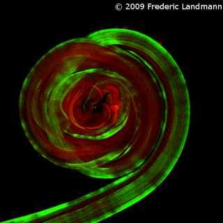
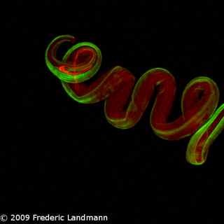
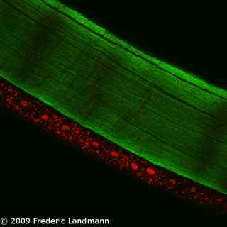
14. Wolbachia in lateral chord (red) with a muscle quadrant (phalloidin, green)
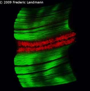
15. Lateral view of previous picture in 3D.
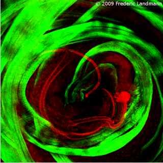
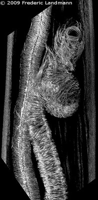
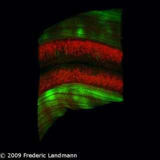
16. in a female (lateral view, 3D)
17. Male and female B. malayi, DAPI staining high resolution in epifluorescence. You can recognize all the parts of the reproductive systems, enjoy!
18. Mature sperm in a B. pahangi male. It seems there are 5 chromosomes.